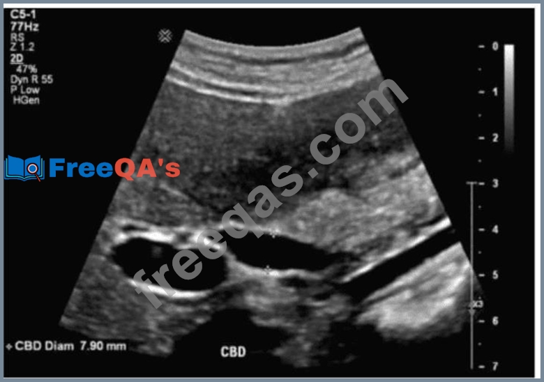
Which term best describes the common bile duct measured in this image of a postcholecystectomy patient?
Correct Answer: A
The ultrasound image shows a measured common bile duct (CBD) diameter of 7.9 mm in a postcholecystectomy patient. In patients who have undergone cholecystectomy, mild dilation of the CBD is considered normal and is a well-recognized post-surgical change. Normal upper limits for CBD diameter: * In patients with a gallbladder: #6 mm is generally considered normal. * In postcholecystectomy patients: up to 10 mm is considered within normal limits, as the CBD compensates for the absence of the gallbladder and slightly enlarges over time. * With aging, the CBD may enlarge by approximately 1 mm per decade after age 60. Therefore, a CBD diameter of 7.9 mm in a patient without a gallbladder is considered normal. Differentiation from other options: * B. Dilated: This would typically refer to a CBD diameter >10 mm in postcholecystectomy patients, or >6 mm in patients with an intact gallbladder. * C. Inflamed: Inflammation refers to wall thickening or hyperemia, which is not evaluated simply by measuring diameter. * D. Atretic: Describes a congenitally absent or severely narrowed duct - not applicable here. References: Rumack CM, Wilson SR, Charboneau JW, Levine D. Diagnostic Ultrasound. 5th Edition. Elsevier, 2018. Chapter: Biliary System, pp. 143-146. American Institute of Ultrasound in Medicine (AIUM) Practice Parameter for the Performance of a Hepatobiliary Ultrasound Examination, 2020. Radiopaedia.org. Common bile duct: https://radiopaedia.org/articles/common-bile-duct
Question 17
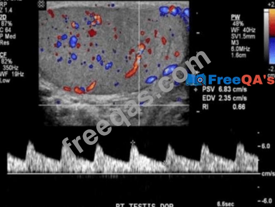
Which best describes the Doppler waveform findings in this image?
Correct Answer: A
The Doppler spectral waveform shown in this image of the right testis demonstrates low-resistance, forward- flowing arterial waveforms with continuous diastolic flow - this is characteristic of normal testicular perfusion. The presence of both color Doppler flow and a resistive index (RI) of 0.66 further supports normal testicular arterial circulation. Key Doppler features of a normal testicular waveform: * Low-resistance waveform (RI typically 0.5-0.75) * Continuous diastolic flow * No reversal of flow or spectral broadening * Color Doppler confirms uniform intratesticular vascularity Clinical context: * Normal testicular flow on Doppler imaging excludes testicular torsion, infarction, or significant inflammation. * Testicular torsion would show either absent or very high-resistance (reduced or absent diastolic flow) waveform. * Epididymo-orchitis may show hyperemia with low resistance but often presents with other gray-scale findings like heterogeneous echotexture or scrotal wall thickening. Differentiation from other options: * B. Increased resistance: RI >0.75 and reduced or reversed diastolic flow; may indicate impending torsion or ischemia. * C. Tardus parvus: A slow systolic upstroke and diminished amplitude; indicates proximal arterial stenosis. * D. Triphasic: Normal waveform in peripheral arteries, such as extremities, not seen in testicular circulation. References: Rumack CM, Wilson SR, Charboneau JW, Levine D. Diagnostic Ultrasound. 5th Edition. Elsevier, 2018. Chapter: Male Pelvis - Testis and Scrotum, pp. 793-800. AIUM Practice Parameter for the Performance of Scrotal Ultrasound Examinations, 2020. Radiopaedia.org. Testicular Doppler assessment: https://radiopaedia.org/articles/testicular-doppler-assessment
Question 18
Which sonographic finding is associated with normal postprocedural Doppler of a transjugular intrahepatic portosystemic shunt (TIPS)?
Correct Answer: D
After successful TIPS placement, the intrahepatic portal venous branches continue to exhibit hepatopetal (toward the liver) flow, while the stent itself shows continuous, relatively high-velocity monophasic flow. Hepatofugal flow in intrahepatic branches may indicate shunt dysfunction. According to Zwiebel's Introduction to Vascular Ultrasound: "Normal post-TIPS Doppler shows hepatopetal flow in the intrahepatic portal veins and continuous high- velocity flow within the stent." Reference: Zwiebel WJ, Pellerito JS. Introduction to Vascular Ultrasound. 6th ed. Elsevier, 2019. AIUM Practice Parameter for the Performance of Portal Venous Ultrasound, 2020. -
Question 19
What is the innermost layer of the gut wall?
Correct Answer: D
The mucosa is the innermost layer of the gastrointestinal wall, consisting of epithelium, lamina propria, and muscularis mucosae. It is responsible for absorption and secretion. The submucosa lies just outside the mucosa. According to Moore's Clinically Oriented Anatomy: "The mucosa is the innermost layer of the gastrointestinal tract, responsible for nutrient absorption and secretion." Reference: Moore KL, Dalley AF, Agur AMR. Clinically Oriented Anatomy. 8th ed. Wolters Kluwer, 2018. Rumack CM, Diagnostic Ultrasound, 5th ed. Elsevier, 2017. - Uploaded image
Question 20
Which sonographic finding distinguishes focal nodular hyperplasia from hepatic adenoma?
Correct Answer: A
The hallmark feature of focal nodular hyperplasia (FNH) is a central stellate scar seen on imaging. This fibrous scar may not always be seen on ultrasound but is a classic distinguishing feature from hepatic adenomas, which usually lack a central scar. According to Rumack's Diagnostic Ultrasound: "Focal nodular hyperplasia often demonstrates a central stellate scar, which may be echogenic or isoechoic." Reference: Rumack CM, Wilson SR, Charboneau JW, Levine D. Diagnostic Ultrasound. 5th ed. Elsevier, 2017. WHO Classification of Digestive System Tumors, 5th ed., IARC, 2019. -