What is the normal Doppler waveform signature of the hepatic veins?
Correct Answer: C
The normal hepatic vein Doppler waveform is triphasic, reflecting cardiac cycle variations in central venous pressure transmitted from the right atrium through the IVC. Loss of triphasicity may suggest elevated right atrial pressures or hepatic venous obstruction. According to Rumack's Diagnostic Ultrasound: "The normal hepatic vein waveform is triphasic due to transmitted right atrial pressure variations." Reference: Rumack CM, Wilson SR, Charboneau JW, Levine D. Diagnostic Ultrasound. 5th ed. Elsevier, 2017. AIUM Practice Parameter for Abdominal Vascular Ultrasound, 2020.
Question 32
Which cause of transudative pleural effusion is most common?
Correct Answer: A
Transudative pleural effusions result from imbalances in hydrostatic and oncotic pressures, most commonly caused by congestive heart failure (CHF). In CHF, elevated hydrostatic pressure in the pulmonary capillaries leads to fluid leakage into the pleural space without significant protein or cellular content (hence, transudate). Exudative effusions (associated with infections, malignancy, and inflammation) are more often seen with pulmonary emboli, lymphoma, or empyema. According to Light's criteria (which differentiates transudates from exudates): "Congestive heart failure remains the leading cause of transudative pleural effusions." (Light RW. Pleural Diseases, 6th ed.). Reference: Light RW. Pleural Diseases. 6th ed. Lippincott Williams & Wilkins, 2013. American Thoracic Society Guidelines for Diagnosis and Management of Pleural Effusion, 2019. -
Question 33
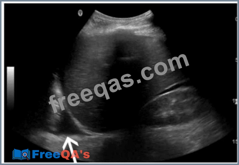
Which finding is indicated by the arrow in this image of the right upper quadrant?
Correct Answer: B
The image provided is a right upper quadrant (RUQ) ultrasound-typically performed during a FAST (Focused Assessment with Sonography in Trauma) exam or for abdominal assessment. The arrow points to an anechoic (black) fluid collection seen above the diaphragm and posterior to the liver. This fluid collection lies within the thoracic cavity, confirming the diagnosis of a pleural effusion. Pleural effusions are seen sonographically as an anechoic or hypoechoic area superior to the diaphragm in the thoracic cavity and often appear triangular or crescent-shaped. The diaphragm is visualized as a curvilinear echogenic structure separating the liver (or spleen) below from the lung space above. Comparison of answer choices: * A. Retroperitoneal hemorrhage would be seen in the posterior abdomen, not above the diaphragm. * B. Pleural effusion is correct-anechoic fluid above the diaphragm is classic for this condition. * C. Mirror image artifact occurs when liver echoes are mirrored across the diaphragm and lung-this is not a mirror artifact. * D. Ascites collects inferior to the diaphragm and around the abdominal organs, not in the thoracic cavity. References: Ma OJ, Mateer JR, Blaivas M. Emergency Ultrasound, 3rd ed. McGraw-Hill; 2014. Moore CL, Copel JA. Point-of-care ultrasonography. N Engl J Med. 2011;364(8):749-757. Rumack CM, Wilson SR, Charboneau JW, Levine D. Diagnostic Ultrasound, 5th ed. Elsevier; 2017.
Question 34
Which sonographic appearance of the normal epididymis is the most common?
Correct Answer: C
The normal epididymis typically appears as a homogeneous structure that is either isoechoic or slightly hypoechoic compared to the testis. The most accurate description is "homogeneous compared to the testis," meaning the texture is uniform. It is not anechoic, nor does it typically show irregular borders unless pathology is present. According to Rumack's Diagnostic Ultrasound: "The normal epididymis appears homogeneous and is isoechoic or slightly hypoechoic relative to the testis." (Rumack CM et al., Diagnostic Ultrasound, 5th ed.) Reference: Rumack CM, Wilson SR, Charboneau JW, Levine D. Diagnostic Ultrasound. 5th ed. Elsevier; 2017. AIUM Practice Parameter for Scrotal Ultrasound, 2020. -
Question 35
Which complication would be associated with retroperitoneal fibrosis?
Correct Answer: D
Retroperitoneal fibrosis can encase and compress the ureters, leading to obstructive uropathy and hydronephrosis. It may also involve other retroperitoneal structures but hydronephrosis is the most common significant complication. According to Rumack's Diagnostic Ultrasound: "Retroperitoneal fibrosis frequently results in ureteral obstruction, leading to hydronephrosis." Reference: Rumack CM, Wilson SR, Charboneau JW, Levine D. Diagnostic Ultrasound. 5th ed. Elsevier, 2017. AIUM Practice Parameter for Abdominal Ultrasound, 2020. -