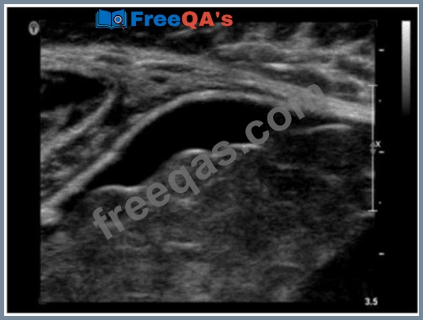
Which description is associated with the normal sonographic appearance of a tendon?
Correct Answer: A
On ultrasound, tendons appear as cord-like hyperechoic structures with linear fibrillar echotexture when imaged in long axis. The fibrils are highly reflective, creating the typical hyperechoic appearance. Posterior shadowing is not typical unless there is calcification. According to Rumack's Diagnostic Ultrasound: "Tendons have a highly organized hyperechoic linear fibrillar pattern when examined along their long axis." Reference: Rumack CM, Wilson SR, Charboneau JW, Levine D. Diagnostic Ultrasound. 5th ed. Elsevier, 2017. AIUM Practice Parameter for Musculoskeletal Ultrasound, 2020. -
Question 37
How are portal veins differentiated from hepatic veins?
Correct Answer: D
Portal veins course horizontally within the liver and have echogenic walls due to their fibrous sheath. In contrast, hepatic veins course vertically toward the IVC and lack echogenic walls. Hepatic veins also demonstrate a triphasic waveform. According to Rumack's Diagnostic Ultrasound: "Portal veins course horizontally within the liver and are easily recognized by their echogenic walls." Reference: Rumack CM, Wilson SR, Charboneau JW, Levine D. Diagnostic Ultrasound. 5th ed. Elsevier, 2017. AIUM Practice Parameter for Liver Ultrasound, 2020. -
Question 38
Which parameter is most likely increased distal to a renal artery stenosis?
Correct Answer: C
Downstream from a significant renal artery stenosis, the acceleration time is prolonged due to delayed systolic upstroke ("tardus-parvus waveform"). This is a sensitive Doppler parameter for detecting hemodynamically significant stenosis. Spectral broadening usually occurs at the stenotic site, not distal to it. According to Zwiebel's Introduction to Vascular Ultrasound: "Prolonged acceleration time and reduced acceleration index characterize tardus-parvus waveforms distal to renal artery stenosis." Reference: Zwiebel WJ, Pellerito JS. Introduction to Vascular Ultrasound. 6th ed. Elsevier, 2019. AIUM Practice Parameter for Renal Artery Duplex Doppler Ultrasound, 2020.
Question 39
Which of the following would optimize visualization of a bladder mass?
Correct Answer: D
A full bladder provides an acoustic window that displaces bowel gas and distends the bladder walls, allowing optimal visualization of any bladder masses or lesions. An empty bladder may collapse, obscuring masses. According to Rumack's Diagnostic Ultrasound: "Bladder evaluation should be performed with the bladder optimally distended to visualize its walls and any intraluminal masses." Reference: Rumack CM, Wilson SR, Charboneau JW, Levine D. Diagnostic Ultrasound. 5th ed. Elsevier, 2017. AIUM Practice Parameter for Bladder Ultrasound, 2020. -
Question 40
Which vascular condition is most likely associated with the sonographic findings demonstrated in this image?
Correct Answer: C
The ultrasound image demonstrates a tubular, anechoic structure coursing anterior to the left portal vein and heading toward the anterior abdominal wall. This is consistent with a recanalized umbilical vein, which is an important collateral pathway that reopens in cases of portal hypertension. Normally, the umbilical vein becomes obliterated after birth and forms the ligamentum teres. However, in the setting of significant portal hypertension, the umbilical vein may recanalize and serve as a collateral route to decompress the portal system. Sonographic features of a recanalized umbilical vein: * Anechoic, tubular structure in the ligamentum teres fissure * Seen anterior to the left portal vein * Color Doppler confirms hepatofugal venous flow * Associated with signs of portal hypertension (e.g., splenomegaly, varices) Differentiation from other options: * A. Budd-Chiari syndrome: Involves hepatic vein outflow obstruction; ultrasound shows absent or narrowed hepatic veins and may have caudate lobe hypertrophy. * B. Splenic artery aneurysm: Typically visualized near the splenic hilum as a pulsatile cystic mass; Doppler shows arterial flow. * D. Median arcuate ligament syndrome: Involves compression of the celiac axis; best assessed with Doppler showing elevated velocities on expiration. References: Rumack CM, Wilson SR, Charboneau JW, Levine D. Diagnostic Ultrasound. 5th Edition. Elsevier, 2018. Chapter: Portal Hypertension and Collaterals, pp. 101-104. American Institute of Ultrasound in Medicine (AIUM). Practice Parameter for the Performance of a Vascular Ultrasound Examination, 2020. Radiopaedia.org. Recanalized umbilical vein: https://radiopaedia.org/articles/recanalised-umbilical-vein